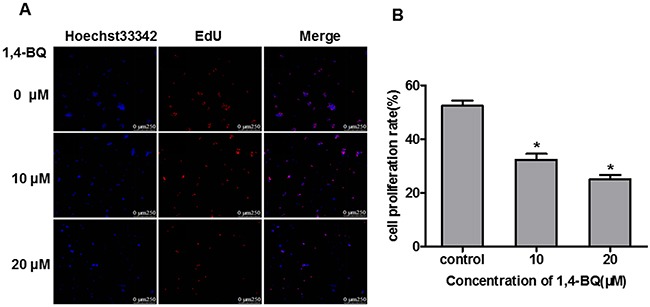
Figure 5. 1, 4-BQ dose-dependently inhibited the proliferation of AHH-1 cells.

(A) The blue fluorescence indicates the nucleus of living cells which were dyed by hoechst33342; The red fluorescence indicates the DNA of cells are being copied and purple is the overlap between the two (The nucleus of living cells are stained blue, and the proliferating cells are stained red by EdU); (B) Cell proliferation rate (%) = Number of proliferating cells / Number of living cells*100%. Results indicated that 1, 4-BQ dose-dependently inhibited the proliferation of AHH-1 cells. *P<0.05, compared with control. Data are expressed as means ± S.D. n=3.
