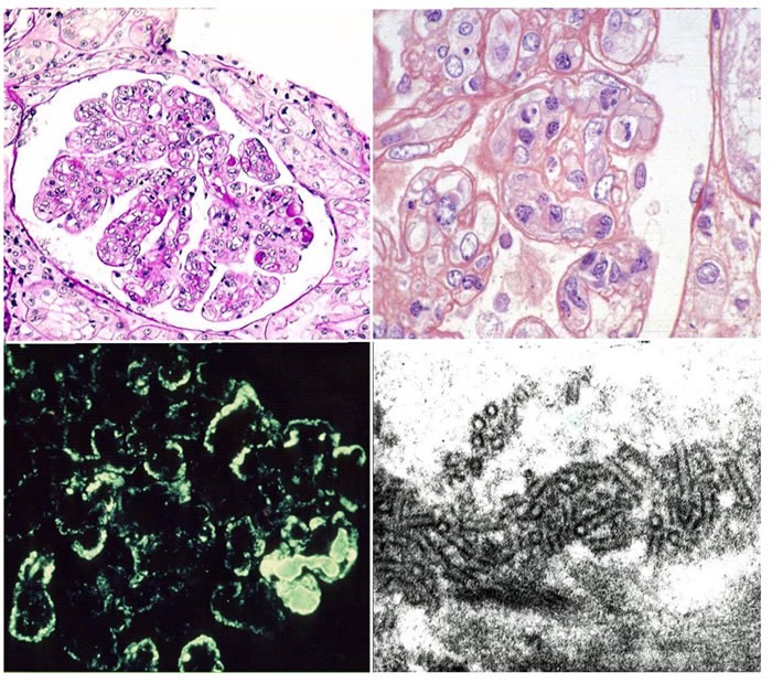
Figure 1. This picture shows the main features of cryoglobulinemic nephritis.

Light microscopy: Upper left side membranoproliferative pattern. Many loops contain pale eosinophilic material consistent with cryoglobulins. Upper right side higher power magnification showing double contour formation. Immunofluorescence (lower left side): subendothelial and mesangial deposition of immune reactants. Electron Microscopy (lower right side): structured appearance of electron dense deposits.
