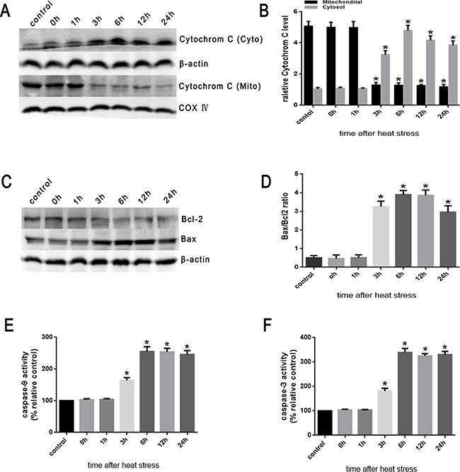
Figure 6. Heat stress induces the activation of mitochondrion-associated pro-apoptotic proteins in SW480 Cells.

SW480 cells underwent intense heat stress (43°C) for 2h and were further incubated at 37°C for different times as indicated (0h, 1h, 3h, 6h, 12h or 24h). A. Intracellular location of cytochrome C was determined by Western blot. β-actin was run as an internal control. COX IV was used as a mitochondrial loading control. B. Quantification of Western blots for cytochrome C after heat stress. C. Bcl-2 and Bax were determined by Western blots. β-actin was run as an internal control. D. Quantification of Western blots for Bax/Bcl-2 ratio after heat stress. E. Enzymatic activity of caspase-9 was measured in cell lysates using a fluorogenic substrate, Ac-LEHD-AFC. F. Enzymatic activity of caspase-3 was measured in cell lysates using a fluorogenic substrate, Ac-DEVD-AMC. The data shown represent the mean ±SD of at least three independent experiments, performed in triplicate. *P < 0.05, statistically significant relative to control (37°C).
