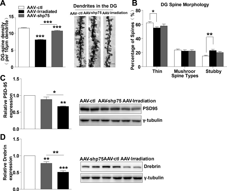
Figure 5. Normalization of p75NTR levels in irradiated rats prevents dendritic spines and synapse-related proteins deficits.
(A) Quantitative analysis showing dendritic spine density in DG region. Right: Representative dendrites of DG granule neurons from AAV-ctl, AAV-irradiation, and AAV-shp75 rats after virus-injection 1 month. (B) Quantified spine types of dendritic spine including thin, mushroom, and stubby morphologies in DG from AAV-ctl, AAV-irradiation, and AAV-shp75 rats after virus-injection 1 month. Western blot for PSD-95 (C) and Drebrin (D) in total hippocampus extracts from AAV-ctl, AAV-irradiation, and AAV-shp75 rats after virus-injection 1 month. All histograms represent mean ± SEM.*p<0.05; **p<0.01; ***p<0.001. n=3-5/group.

