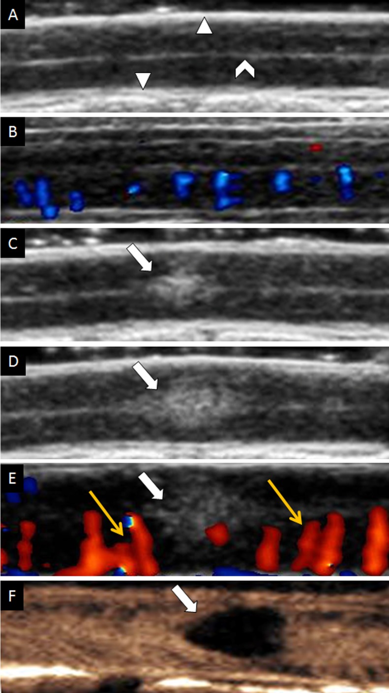
Figure 2. Ultrasound imaging of spinal cord on longitudinal view.
(A) Gray scale of uninjured spinal cord. (B) CDFI of uninjured spinal cord. (C) Contusive spinal cord instantly on gray-scale sonography. (D) Increasing size of contusive spinal cord on gray-scale sonography. (E) CDFI of injured spinal cord. Yellow arrows indicated dilated vessels. (F) CEUS showed the contusive lesion was non-perfusion. The white triangle indicated dura mater of spinal cord; The white caret indicated cerebral aqueduct. The white arrow indicated contusive lesion.

