
Figure 5. PDE3A-ir in most human GIST irrespective of the histological subtype.
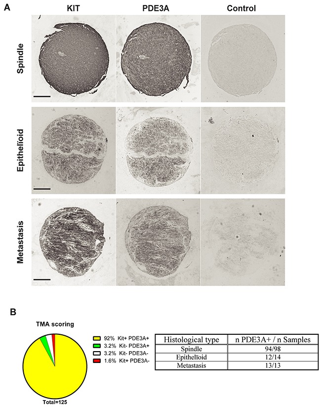
(A) Immunohistochemistry. Examples of hPDE3A-ir in spindle shape, epithelïoid and metastatic human GIST. Widefield microscopy. Scale bar = 200μm. (B) Strong correlation (P-value = 0.0001 (Fisher's exact test)) between PDE3A-ir and KIT-ir in a pool of human GIST TMA. (See Supplementary Tables 4 and 5 for details).