
Figure 6. PDE3A expression in the human GIST882 cell line is modulated by KIT and MEK/ERK inhibition.
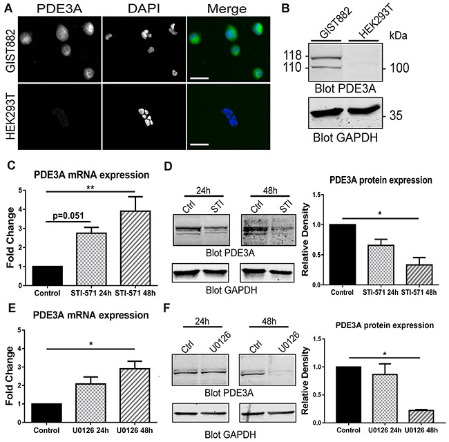
(A) Immunofluorescence. hPDE3A-ir and DAPI nuclear counterstain in GIST882 and HEK293T cells. hPDE3A-ir was detected in GIST882 while no signal was detected in HEK293T. Widefield microscopy. Scale bar = 50μm. (B) Western blot of GIST882 and HEK293T cells probed with anti-hPDE3A and anti-GAPDH as loading control. Bands at 118 kDa and 110 kDa were observed in GIST882 lanes while no band was present in HEK293T lanes. 50μg protein/lane. (C) qPCR of GIST882 cells treated with 1μM of the KIT inhibitor STI-571 for 24H and 48h. PDE3A mRNA expression increased significantly after 48h KIT inhibition. (D) Left panel: Western blot of GIST882 cells treated for 24h and 48h with 1μM STI-571 probed with anti-hPDE3A and anti-GAPDH antibodies. Right panel: Quantification of PDE3A normalized to loading control GAPDH. PDE3A protein expression was significantly reduced after 48h KIT inhibition. 50μg protein/lane. (E) qPCR of GIST882 cells treated with 10 μM of the MEK/ERK inhibitor U0126 for 24h and 48h. PDE3A mRNA expression increased significantly after 48h MEK inhibition. (F) Left panel: Western blot of GIST882 cells treated for 24h and 48h with 10μM U0126 probed with anti-hPDE3A and anti-GAPDH antibodies. Right panel: Quantification of PDE3A normalized to loading control GAPDH. PDE3A protein expression was significantly reduced after 48h MEK/ERK inhibition. 100μg protein/lane. Data presented as mean+/- SEM. P-values (Kruskal-Wallis followed by Dunn's test). *: p {less than or equal to} 0.05, **: p {less than or equal to} 0.01.