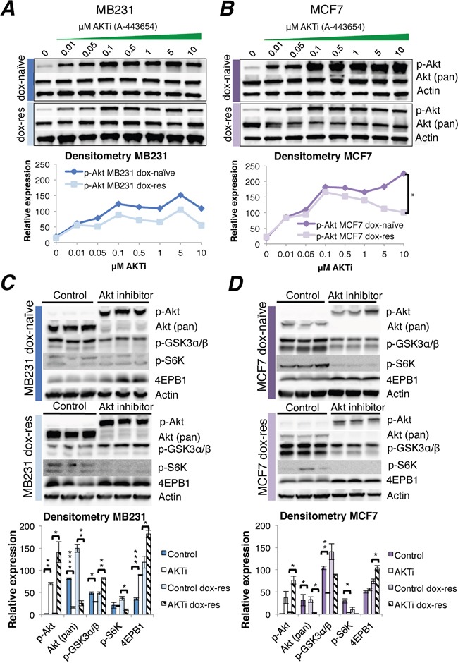
Figure 2. Akt inhibitor treatment of doxorubicin-naïve and doxorubicin-resistant human breast cancer cell lines.

(A-B) Western blots of Akt phosphorylation induced by increasing doses of the Akt inhibitor A-443654, 0-10 μM, 2 hrs exposure in doxorubicin-naïve or doxorubicin-resistant MB231 (A) and MCF7 (B) human breast cancer cells in vitro. Whole cell lysate, 30 μg protein loaded per lane. Densitometries for western blots (A-B) depict the relative protein expression, normalized to actin and total Akt. Phosphorylated Akt increased significantly in doxorubicin-naïve (dox-naïve), compared to doxorubicin-resistant MCF7 cells (dox-res), at AKTi concentrations above 0.5 μM. *p<0.05. (C-D) Western blot analysis of Akt and downstream signaling in doxorubicin-naïve or doxorubicin-resistant MB231 (C) and MCF7 (D) human breast cancer cells, after 24 hrs exposure to A-443654 (MB231: 1 μM, MCF7 0.5 μM) in vitro. Densitometries for western blots (C-D) depict the relative protein expression, normalized to actin. Bars represent the mean protein expression in experiments performed in triplicate ± SEM.
