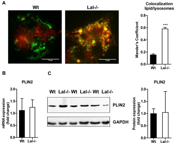
Figure 5. Lysosomal accumulation of lipids in Lal-/- macrophages.

A. Immunofluorescence staining of Wt and Lal-/- macrophages using BODIPY493/503 as neutral lipid dye and Cathepsin D as lysosomal marker. Co-localization analysis was performed using Mander's coefficient (n = 4-5) + SEM. B. mRNA and C. protein expression of the LD marker PLIN2, normalized to the expression of B. cyclophilin and C. GAPDH. Data are shown as means + SD (n = 3-4). ***p ≤ 0.001.
