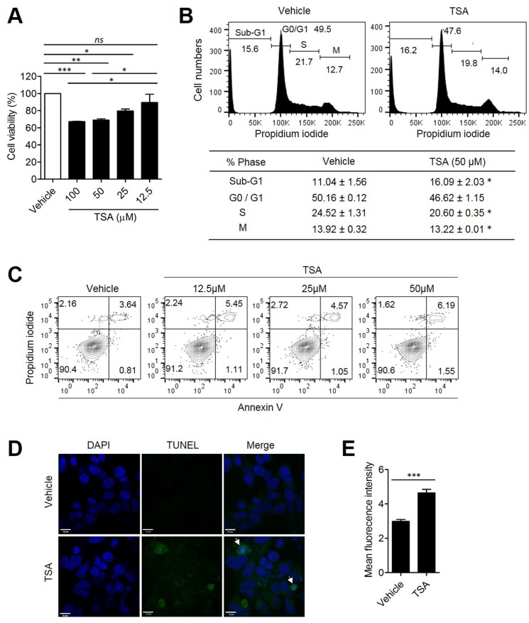
Figure 5. TSA directly induced tumor cell death.
Her2/CT26 cells were treated with the indicated concentration of TSA. (A) Cell viability was evaluated at 24 h after treatment and was determined based on the absorbance at 480 nm (*p<0.05, **p<0.01, and ***p<0.0001, ns; not significant, ANOVA). (B) Propidium iodide-labeled nuclei were analyzed for determination of cell cycle stage. The sub-G1 phase cells containing apoptotic populations were analyzed by flow cytometry (*p<0.01, two-tailed unpaired t-test). (C) Cells were incubated with 0, 12.5, 25, and 50 μM TSA for 24 h. Annexin-V- and propidium iodide-stained cells were analyzed for cell death using flow cytometry. (D) Her2/CT26 cells were treated with 100 μM TSA for 24 h and then stained using the TUNEL assay. Apoptotic cells were examined by confocal microscopy. (E) The mean green fluorescence intensity of TUNEL positive cells is summarized (***p<0.001, two-tailed unpaired t-test).

