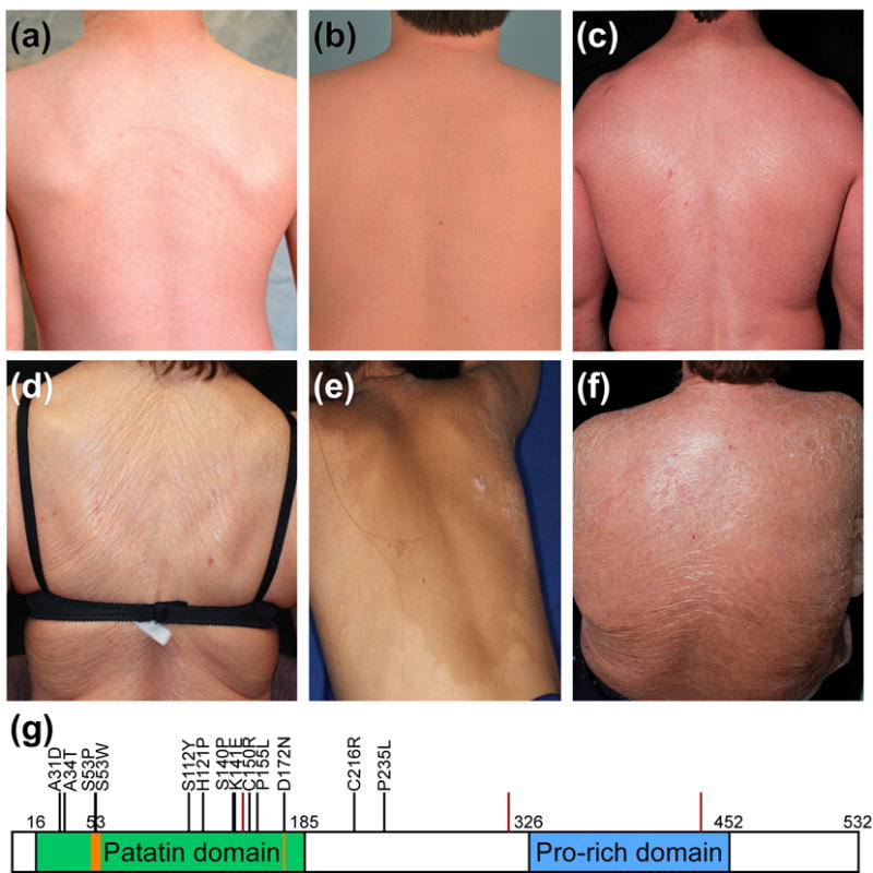
Figure 1. Spectrum of cutaneous phenotypes and PNPLA1 mutation sites in subjects with ichthyosis.

Extent and severity of erythema and scale vary significantly and include: (a) ICH136-1 and (b) ICH431-1, mild erythema and fine white scale; (c) ICH201-4, moderate-severe erythema and fine white scale; (d) ICH162-2 and (e) ICH561-1, minimal erythema and plate-like scale; and (f) ICH201-1, moderate-to-severe erythema with plate-like scale. (g) PNPLA1 protein domains: patatin domain (green), lipid hydrolase catalytic dyad (orange), and proline-rich domain (blue) are indicated; numbers specify amino acid position. Locations of mutations reported herein are shown with black bars and the amino acid change (missense mutations) or red bars (splice site and frameshift mutations).
