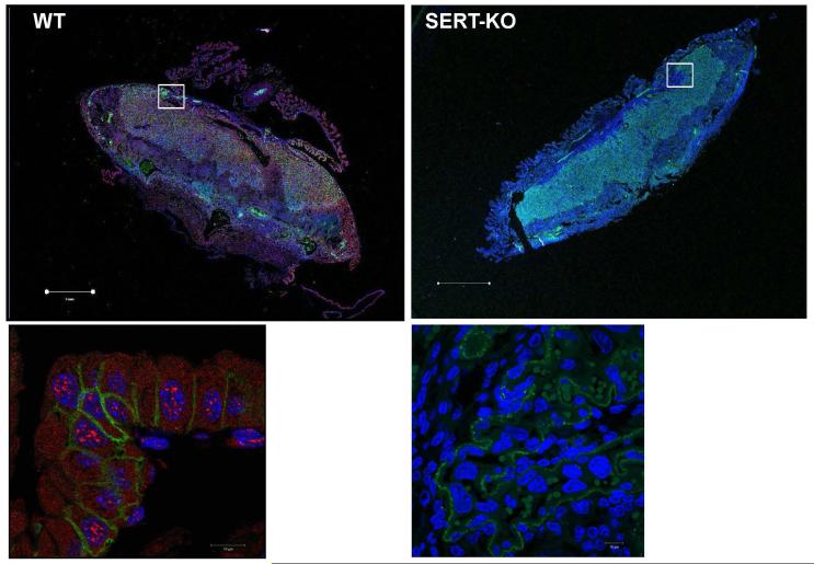
Figure 1. Expression of SERT in E18 mouse placentas.
The 4 μm sections of parafine embeded placentas dissected from E18 WT and SERT-KO mice were dewaxed, rehydrated and subjected to antigen retrieval. Sections were stained as described in the Methods with polyclonal SERT- and monoclonal TROP-1 antibodies (1:100 dilution) followed by Alexa549-conjugated donkey anti-goat and Alexa488 donkey anti-rabbit dye (1:250 dilution). Nuclei were counterstained with DAPI in blue (1:250 dilution). In placentas dissected from SERT-KO mouse only green TROP-1 and blue DAPI staining are found. In WT placentas red, SERT-staining appeared over TROP-1. Cells were analyzed with a Zeiss LSM510 laser confocal microscope. The lower panels are 10X the magnification of the area framed in white in the upper panels. The scale bars for the upper panels are in 1 μm and for the lower panels 10 μm indicate the magnification of the images. Figures show representative images from at least 2 separate experiments.

