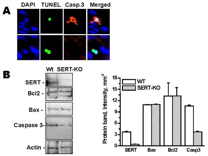
Figure 6. Caspase3 staining of the placentas from SERT-KO mice.
(A) E.18d placentas from WT and SERT-KO mice were first analyzed in TUNEL assays and the same field was evaluated for caspase3 staining (n=3). Only one of the three SERT-KO placentas gave a negligible levels of Caspase3 staining suggest that in the absence of SERT the placental damage occurs in caspase3-independent signaling pathway. The images in each panel are acquired at 400X magnification. The scale bars represent 10 μm. (B) Placental cells prepared from WT and SERT-KO (E.18d) (Yi et al., 2014) were analyzed in 5-12% gradient SDS. The WB analysis were performed with antibodies against Bax or Bcl2 or Caspase-3. The results of Western blot analysis are the summaries of combined data from four densitometric scans of the protein bands.

