Figure 5.

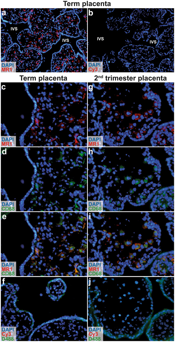
MR1 is expressed by macrophages in foetal villi, but not in syncytiotrophoblasts. Immunofluorescence microscopic images of term placental tissue showing cross sections of chorionic villi and intervillous space (IVS) stained with (a) MR1 (red) or (b) secondary antibody only (control) at magnification × 10. MR1 was expressed by cells inside the villi, but not in syncytiotrophoblasts. Sections of placental villi at (c–e) term and (g–i) gestation week 13 were stained for MR1 (red) and CD68 (green) (×20). (e and i) Merged images showed that MR1 and CD68 were coexpressed by cells in the villi. Sections stained only with secondary antibody (Cy3) and DyLight488-conjugated (D488) streptavidin complex were used as negative controls (f and j) (×20). Nuclei were stained with DAPI (blue). The same microscopy settings were kept for the samples and the corresponding controls, n = 2 for term placentas and n = 1 for 13 week gestation.
