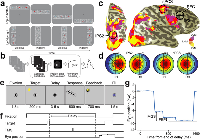
Figure 1.

Experimental procedures. (a) Discrimination task used for topographic mapping. Subjects fixated centrally while covertly attending to a bar consisting of three apertures of moving dots that swept across the screen. Subjects pressed a button to indicate which flanking aperture (left or right; above or below) contained dots moving in the same direction as the center sample aperture. Staircases adjusted dot motion coherence in the flanking apertures to constantly tax attention. White outlines around each aperture were not visible to the subject, but are shown here for clarity. (b) Nonlinear population receptive field (pRF) modeling schematic. Stimulus sequences from the discrimination task were converted into 2D binary contrast apertures and projected onto an isotropic 2D Gaussian that represented a predicted pRF. Static nonlinearity was applied to account for compressive spatial summation. (c) Stimulation sites projected onto the inflated cortical surface of a sample subject. Color indicates the best-fit phase angle by the pRF model. sPCS and IPS2 stimulation sites were localized by our mapping procedure, and dorsolateral PFC stimulation targets were defined by individual subject anatomical landmarks. (d) Visual field map coverage in IPS2 and sPCS. While visual field maps in both regions primarily represent the contralateral hemifield, they also represent portions of the ipsilateral hemifield. (e) Memory-guided saccade task. While fixating, subjects maintained the position of a brief visual target over a memory delay, and then made a saccade to the remembered target. The correct target location was again presented for feedback. Dotted circles depict gaze, but were not visible to subjects. (f) Temporal structure of the experimental task and application of TMS. In order to disrupt the maintenance of information, we applied a short burst of patterned TMS to IPS2, sPCS, or dorsolateral PFC during the middle of the delay period on each trial. (g) An example horizontal eye position trace shows two distinct types of memory error relative to the true target location: the endpoint of an initial memory-guided saccade (MGS), and the final eye position (FEP) following quick corrective saccades (in this example only one) prior to the re-presentation of the target feedback (gray line).
