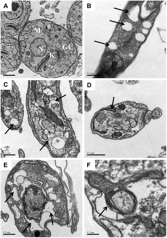
Figure 5.

Transmission electron micrographs of trypomastigotes treated or not with DMS for 24 h. (A) Untreated trypomastigotes presenting a typical morphology of the nucleus (N), kinetoplast (K), mitochondria (M) and Golgi complex (GC). (B,C) Trypomastigotes treated with DMS (1 µM) causes the formation of numerous and atypical vacuoles within the cytoplasm accompanied by a large loss of density. (D,E) Trypomastigotes treated with DMS (2 µM) shows degeneration of mitochondria and intense vacuolization. (F) Trypomastigotes treated with DMS (4 µM) shows myelin-figures. Black arrows indicate alterations cited. Scale bars: A = 1 µm; B–E = 0.5 µM; F = 0.2 µm.
