
Figure 6.
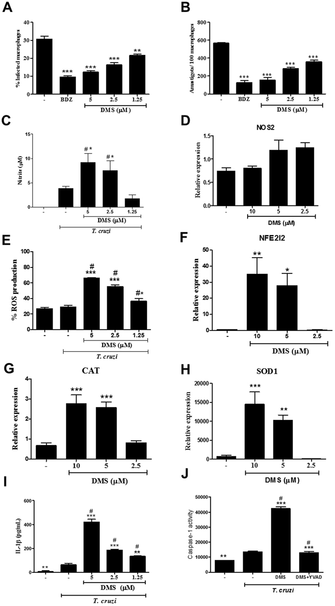
DMS inhibits amastigote proliferation in T. cruzi-infected macrophages, increases NO and ROS and activates caspase 1. The percentage of infected macrophages (A) and the relative number of amastigotes per 100 macrophages (B) were determined by counting hematoxylin and eosin-stained cultures after 72 hours of treatment. (C) Nitric oxide was determined by Griess method after 72 hours of treatment. (D) Relative expression of NOS2 gene in infected macrophages treated or not with DMS. (E) Reactive oxygen species was quantified by stained with 2′,7′-dichlorofluorescin diacetate after 30 minutes of treatment. (F–H) Relative expression of NFE2I2, CAT and SOD1 genes in infected macrophages treated or not with DMS. (I) IL-1β production quantified by ELISA. (J) Caspase-1 activity measured using caspase-Glo 1 inflammasome assay in cultures incubated with complete medium alone, with DMS (5 μM) or with DMS and YVAD (a caspase-1 inhibitor) in triplicate for 2 h. Values represent means ± SEM of 4 determinations. ***P < 0.001; **P < 0.01; *P < 0.05 compared to infected and untreated group; # P < 0.05 compared to uninfected and untreated group.