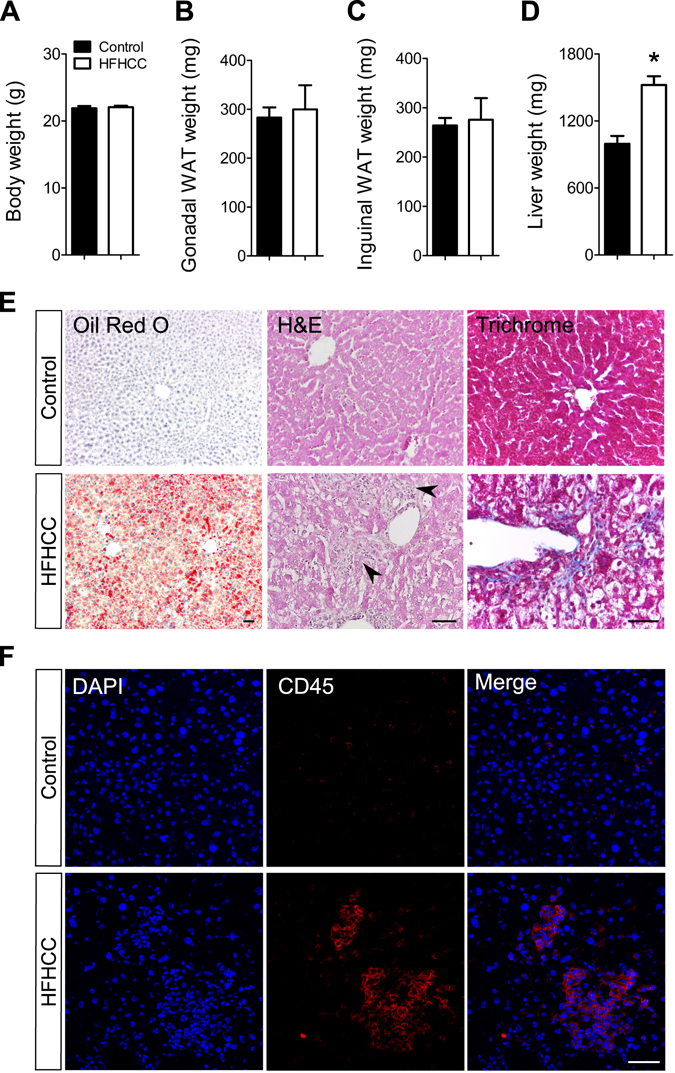
Figure 1.

Weight parameters and hepatic pathology in HFHCC diet induced non-obese NAFLD/NASH. (A) Body weight was not significantly different between mice fed the HFHCC diet for three weeks compared to controls. (B,C) Weights of gonadal and inguinal white adipose tissues (WAT) were not significantly different between mice fed the HFHCC diet compared to controls. (D) Weights of livers from mice fed the HFHCC diet were significantly higher than controls (*p < 0.05). (E) Representative images showing neutral lipid staining using Oil Red O showed significant lipid accumulation (in red) only in the liver of HFHCC diet-fed mice. Histopathology examined after hematoxylin and eosin staining of liver sections showed prominent inflammatory loci (black arrows) only in the livers of HFHCC diet-fed mice. Trichrome staining detected collagen deposits (blue color) only in the livers of HFHCC diet-fed mice. (F) Immunohistochemical staining showed prevalence of CD45(+) immune cells in the livers of HFHCC diet-fed mice but not the control diet-fed mice. DAPI stained for nucleus. Scale bar: 50 µm (For all panels: n = 8 mice on Control diet and n = 6 mice on HFHCC diet were examined).
