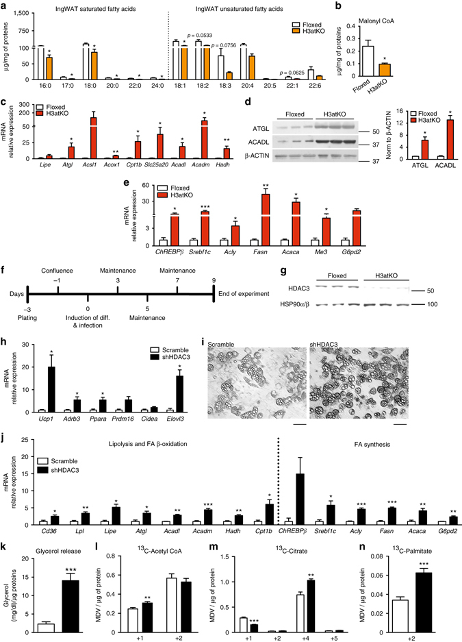
Fig. 4.

Hdac3 ablation activates a futile cycle of FA metabolism in adipocytes. a IngWAT fatty acid quantification by mass spectrometry in floxed and H3atKO mice (n = 7–8 per group); b Malonyl CoA levels in IngWAT of floxed and H3atKO mice (n = 7–8 per group); c Expression of lipolysis and FA β-oxidation genes in IngWAT of floxed and H3atKO mice (n = 9–11 per group); d Western blot analysis and quantification of ATGL and ACADL in IngWAT of floxed and H3atKO mice (n = 3 per group); e Expression of lipogenic genes in IngWAT of floxed and H3atKO mice (n = 9–11 per group); expression of Acly gene was the same reported in Fig. 3d; f Experimental paradigm of differentiation and infection of C3H/10T1/2 cells; g Western blot analysis of HDAC3, showing the deletion of the protein in cells infected with adenovirus expressing a shRNA targeted to Hdac3 (shHDAC3) vs. cell infected with a scrambled control shRNA (scramble; n = 3); h Expression of browning genes in scramble and shHDAC3 infected cells (n = 4 biological replicates per group); i Morphology of scramble and shHDAC3 infected cells, scale bar is 50 μm.; j Expression of lipolytic and lipogenic genes in scramble and shHDAC3 infected cells (n = 4 biological replicates per group); k Glycerol release in scramble and shHDAC3 infected cells (n = 10 per group); l–n 13C-labeled acetyl-CoA, citrate, and palmitate in scramble and shHDAC3 infected cells (n = 6 per group). Data are presented as mean ± s.e.m. Statistical analysis: Student’s t-test, *p < 0.05, **p < 0.01, and ***p < 0.001
