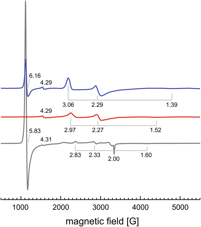
Figure 4.

EPR analysis of LcpK30. Continuous-wave EPR spectra were recorded for LcpK30 as isolated (blue), LcpK30 with imidazole (1 mM, red) and the LcpK30 Lys167Ala variant (grey). Three discernible signals are a broad near-axial signal originating from a six-coordinate low-spin haem, most likely with Lys167 coordinating the haem iron. A signal at g~6 represents a second population of five-coordinate high-spin haem. Added imidazole binds to the haem group, replacing Lys167 and shifting the protein to a low-spin state. The high-spin signal disappears, and the g-values of the low-spin signal shift to 1.52, 2.27 and 2.97, representing the binding of imidazole, with the total population of paramagnetic Fe3+ being reduced.
