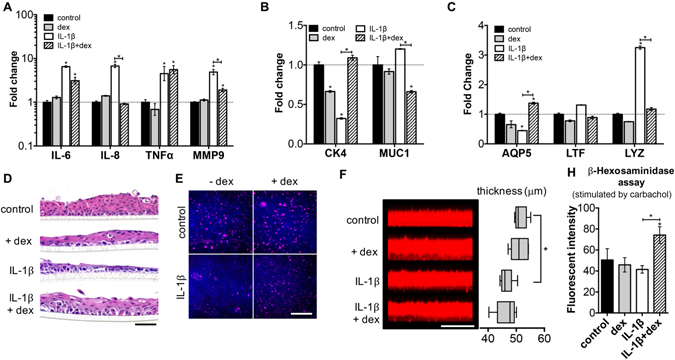
Figure 6.

Responses of direct cell contact model after cytokine IL-1β stimulation. (A–C) Gene expression of the coculture system after the addition of IL-1β. (A) Inflammatory genes; (B) conjunctival epithelial specific genes; (C) lacrimal gland specific genes. (D) The change of epithelial thickness and morphology (H&E staining) after IL-1β exposure. Scale bar 100 μm. (E) MUC5AC staining of the airlifted CECs under various conditions (top view). Scale bar 200 μm. (F) Mucin layer secreted by airlifted CECs imaged by confocal z-scanning. The thickness values were measured using ImageJ. Scale bar 100 μm. (H) β-hexosaminidase assay (supernatant of LG cell spheroids culture; measured by fluorescent intensity). Dexamethasone: dexamethasone, used as a treatment to inflammatory caused by IL-1β. *p < 0.05; **p < 0.001. Significance indicates comparison with the control group if not stated otherwise.
