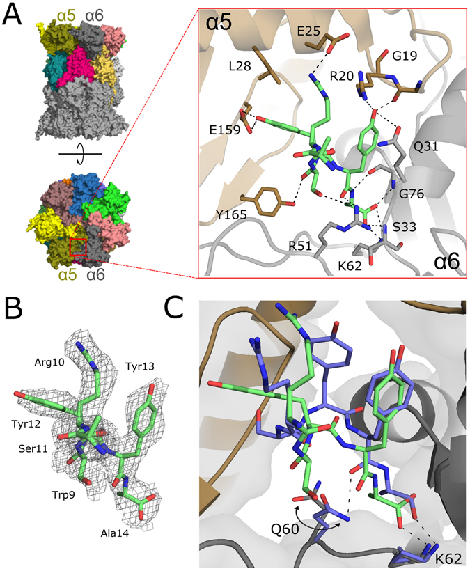
Figure 3.

Interaction of Blm-pep with yeast 20S proteasome. (A) General localization of Blm-pep binding site between subunits α5 and α6 and detailed interactions guiding Blm-pep binding (blow-up). (B) Electron density defining Blm-pep fragment included in the model (2Fo-Fc omit map contoured at 1σ level). (C) Comparison of the binding modes of Blm-pep (green) and Blm10 (blue) at the surface of yeast 20S proteasome (the difference in interaction of both activators with Q60 is highlighted).
