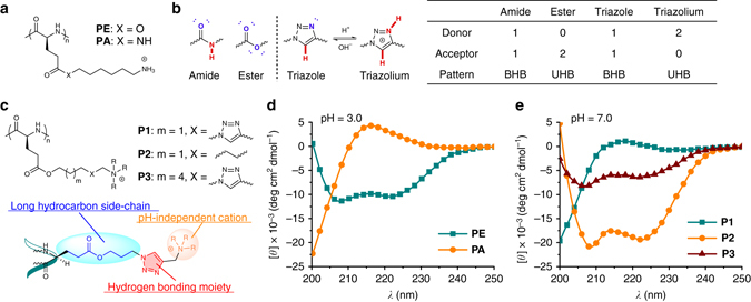
Fig. 2.

Side-chain triazole groups disrupt the backbone α-helical conformation. a Chemical structures of PE and PA. b H-bonding pattern analysis of amide, ester, 1,2,3-triazole, and 1,2,3-triazolium. H-bond donors and acceptors are highlighted in red and blue, respectively. c Chemical structures of P1-P3. The molecular design of triazole polypeptides is highlighted, where each component is essential for the study. d, e CD spectra of polypeptides. PE and PA were analyzed at pH 3.0 (d), and P1-P3 were analyzed at pH 7.0 (e)
