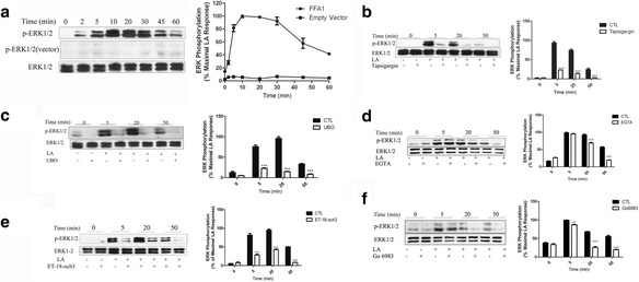
Fig. 1.

Involvement of IP3/Ca2+ and DAG/PKC in FFA1-mediated ERK1/2 activation. a – HEK293 cells expressing FLAG-FFA1 or an empty vector(pCMV-Flag) were starved in serum-free media for 18–24 h followed by a challenge with 10 μM LA for the indicated time periods. ERK1/2 phosphorylation was assessed using western blot as described in the Methods section, and corresponding immunoblots were quantified using the Bio-Rad Quantity One Imaging system. b – Serum-starved FFA1-HEK293 cells were pretreated with DMSO or thapsigargin (0.5 μM) for 1 h, and the cells were then stimulated with 10 μM LA for the indicated time. c – Serum-starved FFA1-HEK293 cells were pretreated with DMSO or UBO-QIC (1 μM) for 1 h, and the cells were then stimulated with 10 μM LA for the indicated time. d – Serum-starved FFA1-HEK293 cells were pretreated with DMSO or calcium chelator EGTA (10 μM) for 1 h, and the cells were then stimulated with 10 μM LA for the indicated time. e – Serum-starved FFA1-HEK293 cells were pretreated with DMSO or phosphoinositide-specific phospholipase C (PI-PLC) inhibitor ET-18-och-3 (10 μM) for 1 h, and the cells were stimulated with 10 μM LA for the indicated time. f – To investigate the role of PKC, serum-starved FFA1-HEK293 cells were pretreated with DMSO or Go6983 (10 μM) for 1 h, and then stimulated with 10 μM LA for the indicated time. Error bars represent the SEM for three replicates. The data shown are representative of at least three replicate independent experiments. Data were analyzed using Student’s t-test. **p < 0.001; ***p < 0.001
