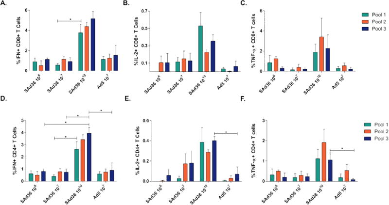
Figure 4. Cytokine production following ex vivo stimulation with PyCMP peptide pools.

Female CB6F1/J mice (n = 5 per group) were immunized according to the regimens described in Table 1. Splenocytes were obtained five days after the final immunization and incubated with peptide pools containing pools of 15AA overlapping peptides representing PyCMP. Following 6 hours of stimulation, cells were intracellularly stained and processed for flow cytometry. Results are presented after background subtraction. Percentage of CD8+ T cells capable of producing IFN-γ (A), IL-2 (B), and TNF-α (C) after stimulation. Percentage of CD4+ T cells capable of producing IFN-γ (D), IL-2 (E), and TNF-α (F) following stimulation. Statistical analysis was conducted using Kruskal-Wallis test to determine differences between the immunization regimens. Statistically significant differences (p < 0.05) are indicated by a single asterisk.
