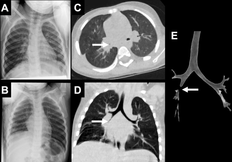
Fig. 4.

2-year-old boy with cough and exertional wheeze underwent frontal (not shown), left lateral (A) and right lateral decubitus (B) radiograph without evidence of foreign body or air trapping, respectively. Ultralow-dose chest CT reconstructed with MBIR (C, D) demonstrates occlusion of the right bronchus intermedius that is also depicted on 3D volumetric reformat of the central airways (E, white arrows). MBIR = Model Based Iterative Reconstruction
