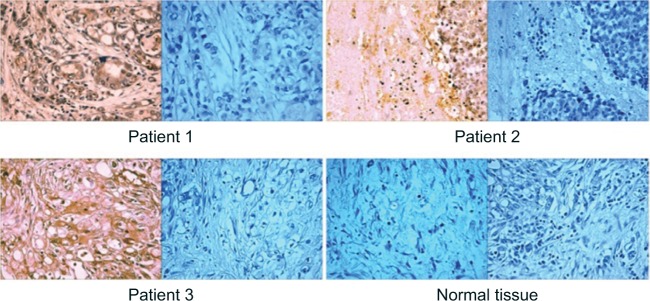
Figure 1.
Imunohistochemical staining of pancreatic cancers (×20) from three patients with positive staining of tumors with GluN2B antibodies (left panels) and negative controls of GluN2B antibodies in the presence of excess peptide antigen (right panels).
Note: Two included normal pancreatic tissues, that give no staining with GluN2B antibodies (last two panels).

