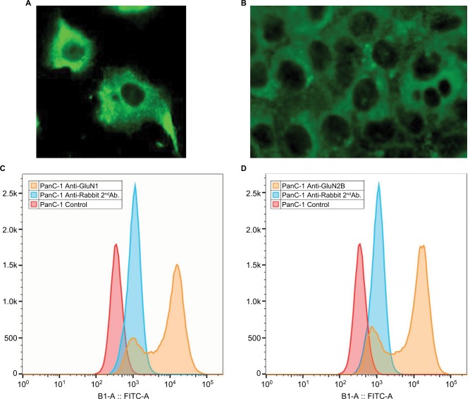
Figure 2.
Demonstration of surface location of GluN1 on pancreatic cancer cells.
Notes: Confocal images with PANN1 antibodies on pancreatic cancer (A) PanC-1 and (B) BXCPC-3 (magnification ×40); Flow cytometry using second Ab fluorescence with (C) GluN1 antibodies, and (D) GluN2B antibodies. Red and blue peaks are negative controls; indications are that ~80% of cells have a positive reaction.
Abbreviation: FITC, fluorescein isothiocyanate.

