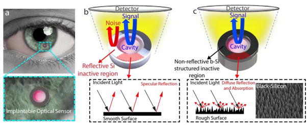
Figure 1. b-Si structured IOP sensor.
(a) Conceptual illustration of the implantable IOP sensor with a microscale optical cavity and a zoomed-in image. (b) A Si-only sensor consisting of an active core cavity and a reflective peripheral inactive region. Reflection arises from both the cavity (shown in blue) and the Si (shown in red). (c) A b-Si sensor with integrated b-Si on the inactive hollow disk. Only the reflection from the core cavity is captured. The inset is a SEM image of the b-Si grown on the inactive region.

