Figure 2.
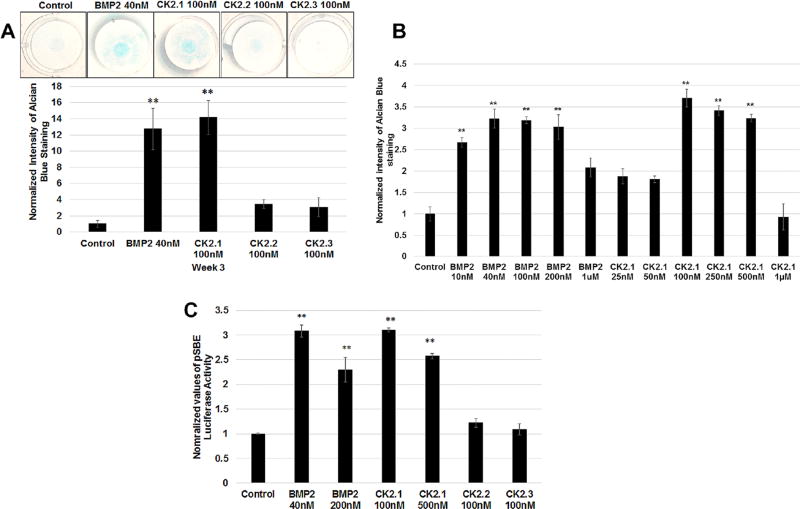
CK2.1 but not CK2.2 or CK2.3 induced chondrogenesis in C3H10T1/2 cells. (A) C3H10T1/2 micromass cultures were treated with either BMP2 (40 nM) or peptides CK2.1 and CK2.2, CK2.3 at (100nM) and stained with Alcian blue for 3 weeks. BMP2-and CK2.1-treated cells showed a significant increase of ECM containing proteoglycans. (B) Concentration curve of micromass stimulated with CK2.1 and BMP2. The treatments identified the concentrations of CK2.1 at 100–500nM as the optimal doses for inducing chondrogenesis. (C) Smad reporter gene assay performed on C3H10T1/2 cells stimulated with CK2.1 at 100nM or 500nM, CK2.2 and CK2.3 at 100 nM, and BMP2 at 40nM or 200 nM. Only CK2.1 and BMP2 induced Smad activity and similar SMAD activity was observed across the equivalent concentrations.

