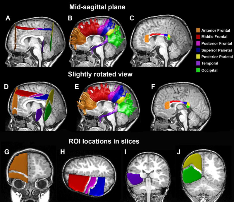
Figure 1.

Fiber tract identification. (A,D) Left ROIs in an individual subject (male, 43 months old, with autism). Homologous ROIs were also defined in the right hemisphere. Tracts were identified by selecting fibers that intersected the left ROI, corpus callosum ROI, and homologous right ROI. (B,E) The seven CC fiber tracts that were identified in this individual. (C,F) Clipped segments of the CC fiber tracts. The same individual is presented in a mid-sagittal view (A–C) and a slightly rotated view (D–F) demonstrating the relative ROI locations in the left hemisphere. Diffusivity measures were extracted from the 10mm mid-sagittal segments presented in C and F. In addition, we present the specific coronal slice containing the anterior frontal ROI (G), the axial slice containing the middle-frontal, posterior frontal, and superior parietal ROIs (H), the coronal slice containing the temporal ROI (I), and the coronal slice containing the posterior parietal and occipital ROIs (J).
