Fig. 1.

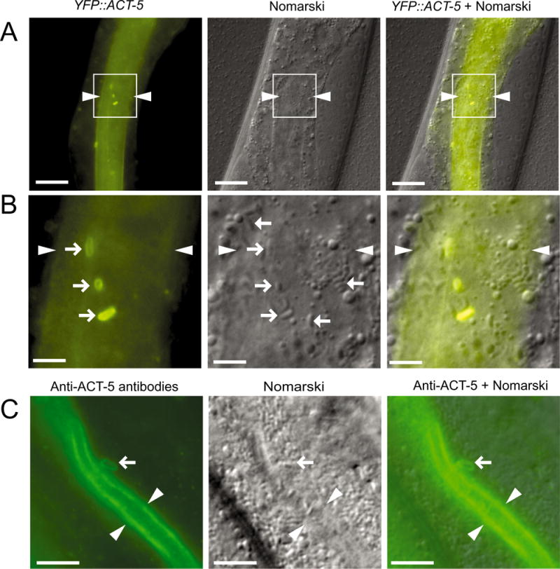
Intracellular N. parisii spores near the lumenal membrane of C. elegans intestinal cells acquire an actin ‘coat’.
A. YFP∷ACT-5 forms ‘coats’ around apically localized spores: region in white box is magnified in B. Arrowheads indicate lumen. Scale bars are 20 μm.
B. Right-pointing arrows indicate apical spores with a YFP∷ACT-5 coat. Left-pointing arrows indicate spores further from the membrane that lack a YFP∷ACT-5 coat. Arrowheads indicate lumen. Scale bars are 5 μm.
C. Anti-ACT-5 antibody staining of infected animals. Arrow indicates spore near lumen with ACT-5 staining similar to those observed with YFP∷ACT-5. Arrowheads indicate lumen. Scale bars are 10 μm.
A–C. Animals imaged at approximately 43 hpi.
