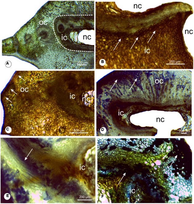
FIGURE 5.

Histochemical analysis of leaflet galls induced by Bystracoccus mataybae (Eriococcidae) on Matayba guianensis (Sapindaceae). (A) Transverse section of a gall with no staining, evidencing the distribution of the chlorophyllous tissue. (B) Histochemical detection of oxygen reactive species (ROS) with DAB (3,3′-diaminobenzidine). The positive reaction is evidenced by the brown spots. (C) Histochemical detection of ROS with DAB in the outer and inner cortex of galls. (D) Proanthocyanidins were detected with DMACA (p-dimethylaminocinnamaldehyde) at the chlorophyllous tissue. (E) Detail of proanthocyanidins in cells around the nymphal chamber, and in lignified cells. (F) Phenolic compounds detected with 2% ferrous sulfate in 10% formalin in gall outer cortical tissues, continuous to the chlorophyllous tissue. ic, inner cortex; nc, nymphal chamber; oc, outer cortex;
