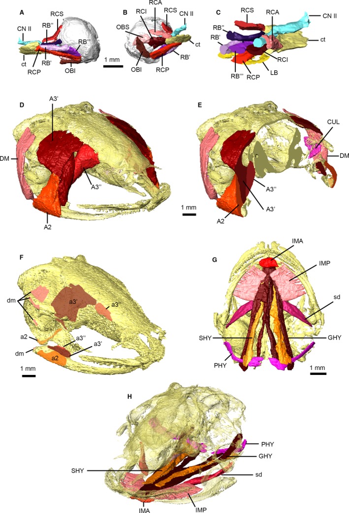
Figure 4.

Head musculature of Xenopus laevis. Musculature of the right orbit in posterolateral (A) and posteromedial (B) views, and in transverse cross‐section through the base of the eyeball (C). Jaw musculature in anterolateral view (D) and transverse cross‐section through the coronoid process (E), and jaw muscle attachments on the skull upper and lower jaws (F). Hyoid musculature in ventral view (G) and posterior oblique view (H) with the skull transparent. Main muscle masses are identified using uppercase abbreviations; muscle attachment sites, small muscle slips and non‐muscle structures are identified using lowercase abbreviations. A2, M. adductor mandibulae A2 (masseter); a2, attachments of A2; A3′, M. adductor mandibular A2 PVM and A3′ (temporalis); a3′, attachments of A3′; A3″, M. adductor mandibulae A3″ (pterygoideus); a3″, attachments of A3″; CN II, optic nerve; ct, central tendon; CUL, M. cucullaris; DM, M. depressor mandibulae; dm, attachments of DM; GHY, M. geniohyoideus; IMA, M. intermandibularis anterior; IMP, M. intermandibularis posterior; LB, M. levator bulbi; PHY, M. petrohyoideus posterior; OBI, M. obliquus inferior; OBS, M. obliquus superior; RB′/RB″/RB′″, portions of M. retractor bulbi; RCA, M. rectus anterior; RCI, M. rectus inferior; RCP, M. rectus posterior; RCS, M. rectus superior; sd, subhyoideus portion of IMP; SHY, M. sternohyoideus.
