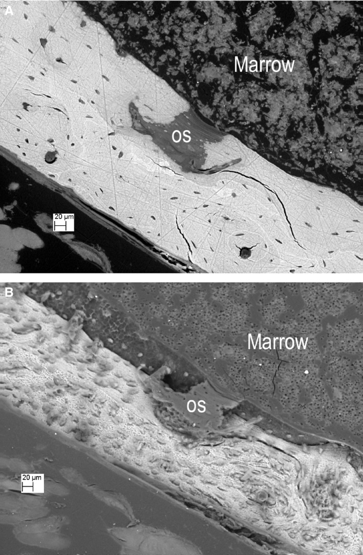
Figure 6.

Same field before and after etching. (A) Knockout (KO) tibia, PMMA‐embedded ammonium triiodide stained to reveal soft tissue components, showing an osteoid patch at the centre. Surface normal to electron beam. [All other scanning electron microscopy (SEM) images in this MS were also recorded at normal electron beam incidence]. (B) The same region after ‘DMG Icon‐Etch’ HCl gel etching for 1 min, followed by 5% NaOCl for 10 min and ammonium triiodide staining. PMMA within the osteoid patch (os) is left proud after the combined etch treatment. Osteocyte lacunae also appear as ‘casts’ above the etched surface. Surface tilted at 33 °.
