
Figure 4.
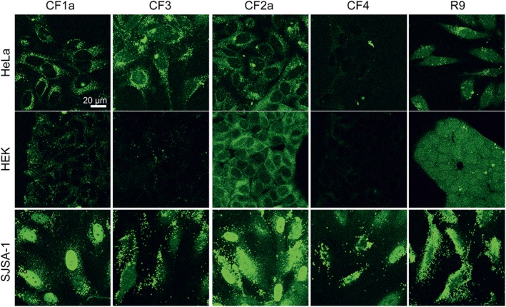
Intracellular distribution of peptides after a 24 h incubation. Cells were incubated with 20 μM of the CF‐labelled peptides in RPMI +10% FCS and washed and confocal images were recorded. R9 was used as a control CPP. The figure shows one representative experiment of three independent repetitions.