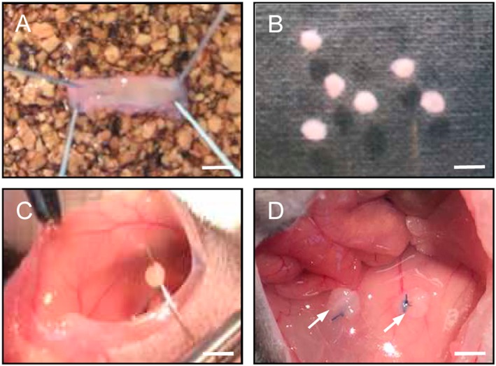
Figure 1.

Surgical induction of murine endometriotic lesions in the peritoneal cavity. Uterine tissue samples (B) were isolated from the longitudinally opened uterine horn (A) of a donor mouse by means of a 2 mm dermal biopsy punch and sutured to the abdominal wall of a syngeneic recipient mouse (C). Typical appearance of the tissue samples directly after fixation (D, arrows). Scale bars: A = 5 mm; B = 2.5 mm; C = 3.3 mm; D = 2 mm.
