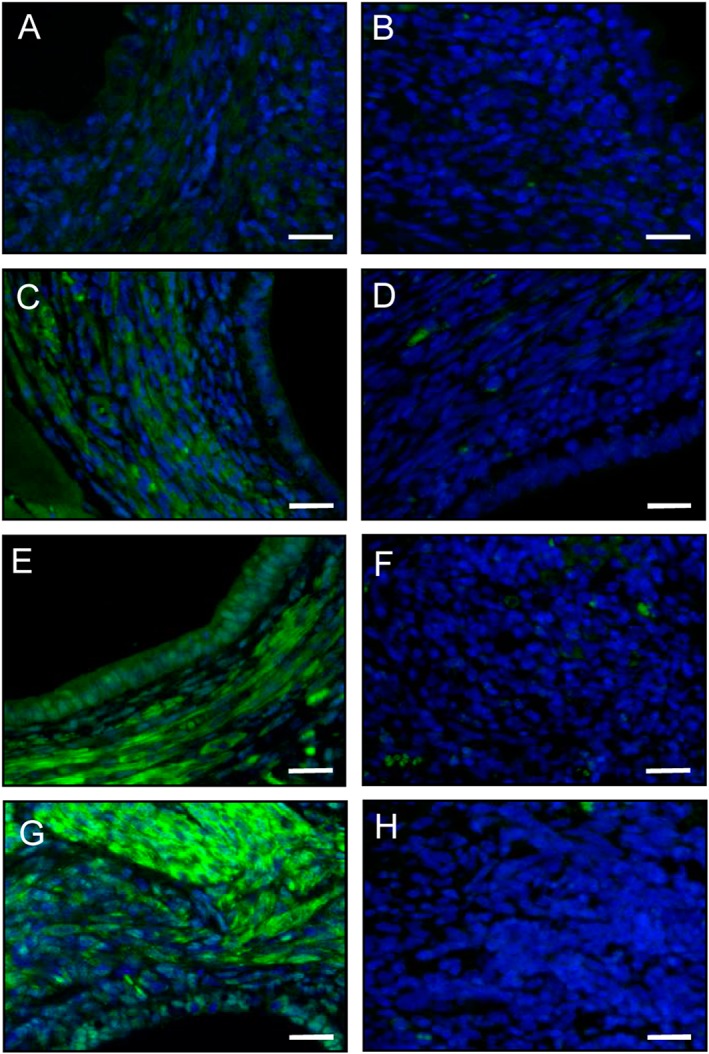
Figure 3.

Immunofluorescence analysis of PPAR‐γ expression within endometriotic lesions. Immunofluorescent detection of PPAR‐γ within endometriotic lesions on day 28 after surgical induction by fixation of uterine tissue samples to the abdominal wall of a vehicle‐treated control (A, B) as well as a parecoxib‐ (C, D), a telmisartan‐ (E, F) and a parecoxib/telmisartan‐ (G, H) treated C57BL/6 mouse. Sections were stained with Hoechst 33342 to identify cell nuclei (blue) and an antibody against PPAR‐γ (green). Sections solely incubated with the secondary antibody served as negative controls (B, D, F, H; green signals = autofluorescence of erythrocytes). Scale bars: 20 μm.
