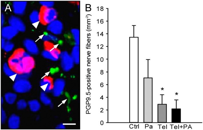
Figure 5.

Immunohistochemical analysis of nerve fibre ingrowth into endometriotic lesions. (A) Immunofluorescent detection of microvessels (arrowheads) and nerve fibres (arrows) within an endometriotic lesion on day 28 after surgical induction by fixation of an uterine tissue sample to the abdominal wall of a vehicle‐treated control mouse. The section was stained with Hoechst 33342 to identify cell nuclei (blue), an antibody against CD31 (red) for the detection of microvessels and an antibody against PGP9.5 (green) for the detection of nerve fibres. Scale bar: 10 μm. (B) Density of PGP9.5‐positive nerve fibres (mm−2) within endometriotic lesions of vehicle‐treated controls (Ctrl), parecoxib‐ (Pa), telmisartan‐ (Tel) and parecoxib/telmisartan‐ (Tel+PA) treated C57BL/6 mice. Means ± SEM (n = 10 for each experimental group). *P < 0.05 versus control.
