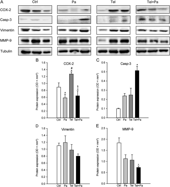
Figure 6.

Western blot analysis of endometriotic lesions. (A) Expression of COX‐2, cleaved caspase‐3 (casp‐3), vimentin, MMP‐9 and tubulin within endometriotic lesions on day 28 after surgical induction by fixation of uterine tissue samples to the abdominal wall of vehicle‐treated control (Ctrl), parecoxib‐ (Pa), telmisartan‐ (Tel) and parecoxib/telmisartan‐treated (Tel + Pa) C57BL/6 mice as assessed by Western blot analyses. Densitometric analyses of expression (OD × mm2) of COX‐2 (B), cleaved caspase‐3 (C), vimentin (D) and MMP‐9 (E) within endometriotic lesions of vehicle‐treated control (Ctrl), parecoxib‐ (Pa), telmisartan‐ (Tel) and parecoxib/telmisartan‐(Tel+Pa) treated C57BL/6 mice. Data were normalized to tubulin signals. Means ± SEM (n = 6 for each experimental group). *P < 0.05 versus control; # P < 0.05 versus parecoxib; § P < 0.05 versus telmisartan.
