
Figure 6.
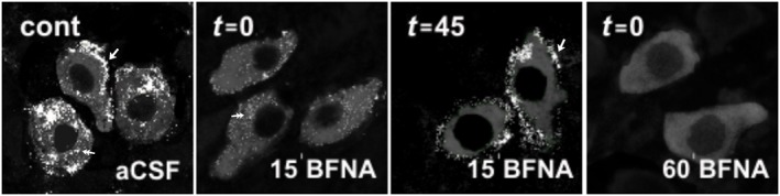
Confocal laser scanning microscopy imaging of μ receptor‐immunoreactivity (ir) in the LC before and after β‐FNA perfusion. In the absence of β‐FNA (control condition, cont), μ receptor‐ir (white) is identified as patches distributed both in the cytoplasm (double arrow) and in association with the plasma membrane (single arrow) in TH‐containing neurons (grey). Immediately after treatment with ß‐FNA (300 nM, 15 min, t 15 = 0), μ receptor‐ir is markedly depleted from the neuronal membrane, but μ receptor‐immunoreactive puncta are still observed within the cytoplasm (double arrow). Forty five min later (t 15 = 45), the μ receptor is present again in the cell membrane (single arrow). Immediately after ß‐FNA (300 nM, 60 min, t 60 = 0) application, μ receptor‐ir is not detected in the cell membrane or the cytoplasmic compartment. All images displayed were acquired with identical laser intensity and detector gain parameters.