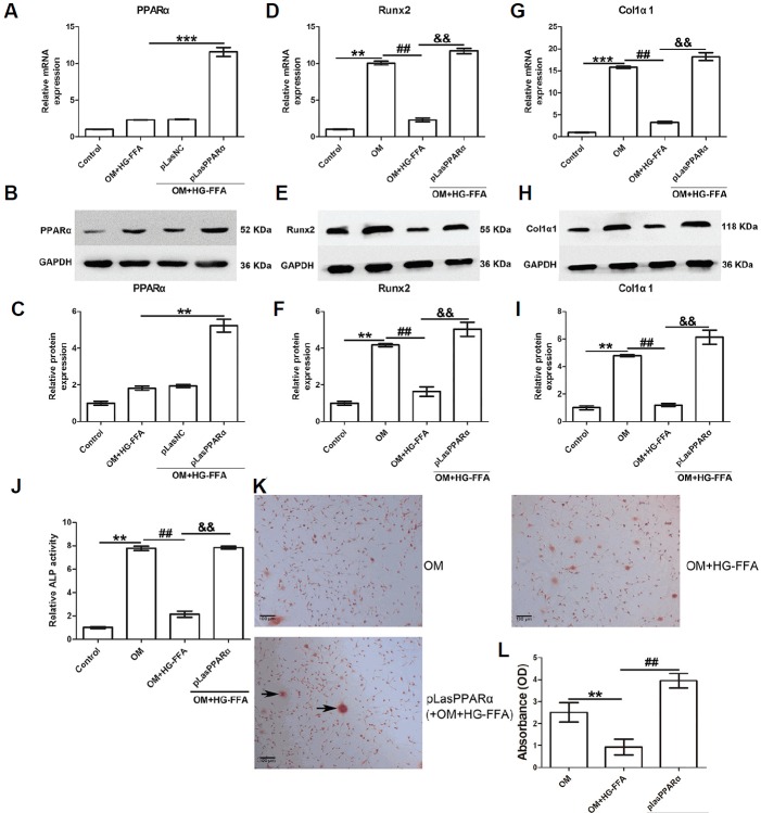
Fig. 2.
PPARα overexpression facilitates osteogenic differentiation.
The expression of PPARα, Runx2, and Col1α1 was evaluated 14 days after osteogenic induction and HG-FFA treatment. (A–C) PPARα was overexpressed through pLasPPARα transfection. PPARα overexpression significantly restored Runx2 (D–F), Col1α1 (G–I) expression, and ALP activity (J) compared to the OM with HG-FFA treatment group. The Alizarin Red staining results demonstrated that PPARα overexpression induced more mineralized nodules (K) and therefore increased absorbance (L) under HG-FFA conditions, compared to the OM with HG-FFA treatment only. (C, F, and I) The densitometry analysis of Western blot results. *** indicates P < 0.001; **, ## and && indicate P < 0.01. N = 3. Scale bars = 100 μm.

