
Figure 4.
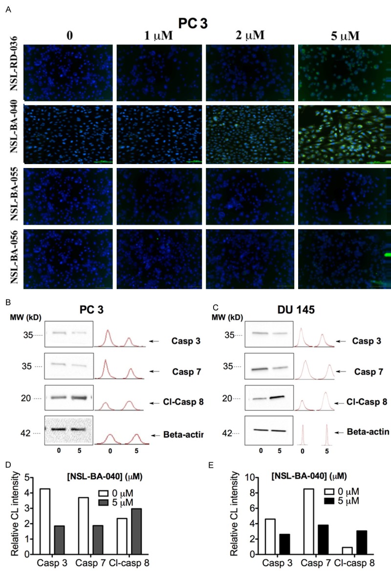
PCAIs-induced apoptosis in PCa cells lines is associated with caspase 3, 7 and 8 activation. A: Caspase 3/7 activation was observed after treating PC 3 cells with 1 to 5 μM PCAIs for 48 h and then reacting with the fluorescent caspase 3/7 irreversible inhibitor; green FLICA. Images were taken with Nikon DS Qi2 digital camera linked to a Nikon Eclipse Ti 100 inverted microscope. B, D: Western blot analysis showing the expression of caspase 3, 7 and cleaved caspase 8 in DU 145 and PC 3 cells after treatment with 5 μM NSL-BA-040. C, E: Bar graph showing the chemiluminescence intensity (CL) of the protein bands relative to control.