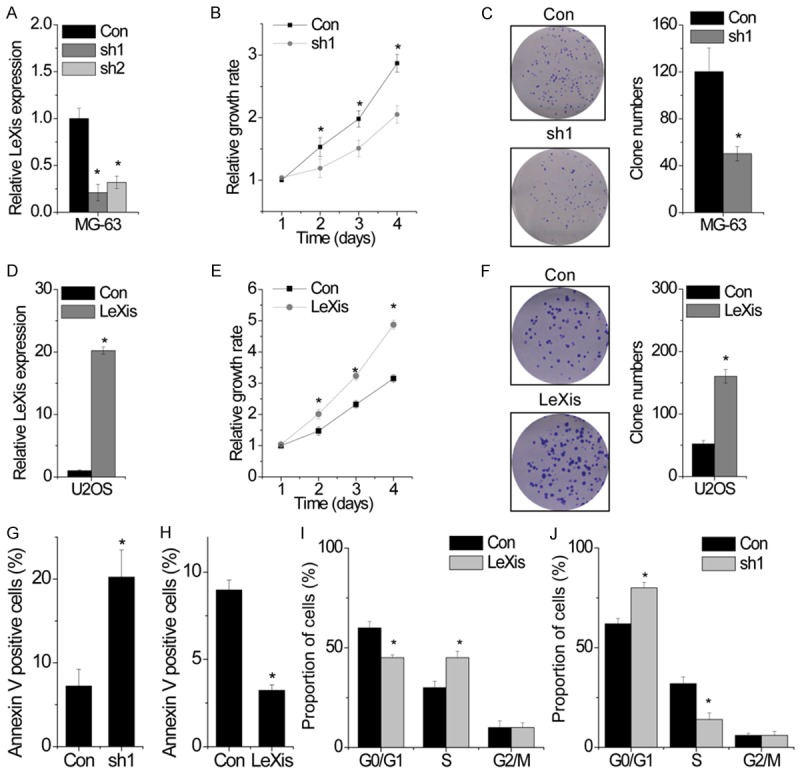
Figure 2.

LeXis enhances OS cell proliferation and inhibits apoptosis in vitro. (A) The relative expression of LeXis in control and LeXis silenced MG-63 cells was detected by qPCR. (B) The cell growth rates were determined by performing CCK-8 assay. Knockdown LeXis in MG-63 cells significantly suppressed cell proliferation, relative to control cells. (C) Colony formation assay of control and LeXis silenced MG-63 cells. Representative graphs are shown. (D) The relative expression of LeXis in control and LeXis overexpressed U2OS cells was detected by qPCR. (E) The cell growth rates were determined by performing CCK-8 assay. Overexpression of LeXis in U2OS cells significantly suppressed cell proliferation, relative to control cells. (F) Colony formation assay of control and LeXis overexpressed U2OS cells. Representative graphs are shown. (G, H) Cells with LeXis knockdown (G) or overexpression (H) were stained with a combination of annexin V and 7-AAD and analyzed by FACS. Cells positive for annexin V staining were counted as apoptotic cells, and the percentage of apoptotic cells is shown. (I, J) FACS analysis showing significant decreases or increases of cells in G0/G1 or S phase, respectively, in U2OS cells with LeXis overexpression (I). In contrast, cells in S phase population were significantly decreased when LeXis was silenced in MG-63 cells (J). Data are shown as mean ± SD; *P < 0.05 (Student’s t test).
