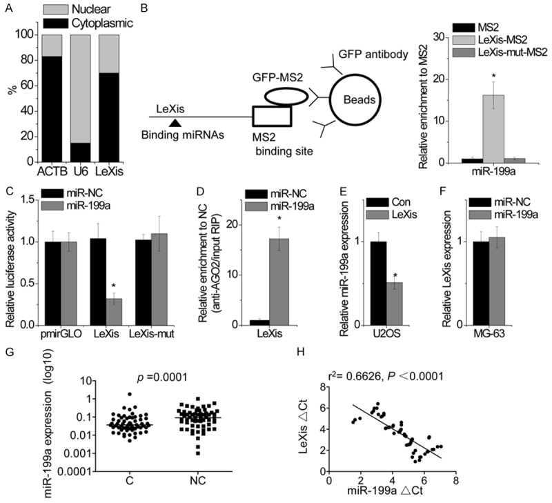
Figure 4.

LeXis directly interacts with miR-199a. A. Representative analysis of LeXis distribution by cellular fractionation of MG-63 cells. ACTB mRNA and U6 RNA served as controls for cytoplasmic and nuclear RNAs, respectively. B. MS2-RIP followed by microRNA qPCR to detect microRNAs endogenously associated with LeXis. C. Luciferase activity in MG-63 cells cotransfected with miR-199a and luciferase reporters containing nothing, LeXis or mutant transcript. Data are presented as the relative ratio of firefly luciferase activity to renilla luciferase activity. D. Anti-AGO2 RIP was performed in MG-63 cells transiently overexpressing miR-199a, followed by qPCR to detect LeXis associated with AGO2. E. The effect of LeXis overexpression on miR-199a expression was detected by qPCR in U2OS cells. F. The effcct of miR-199a overexpression on LeXis expression was determined by qPCR in MG-63 cells. G. Expression of miR-199a was measured in 60 pairs of OS cancerous tissues (C) and adjacent noncancerous bone tissues (NC) using qPCR. H. The correlation between LeXis and miR-199a expression in OS tissues. Data are shown as mean ± SD; *P < 0.05 (Student’s t test).
