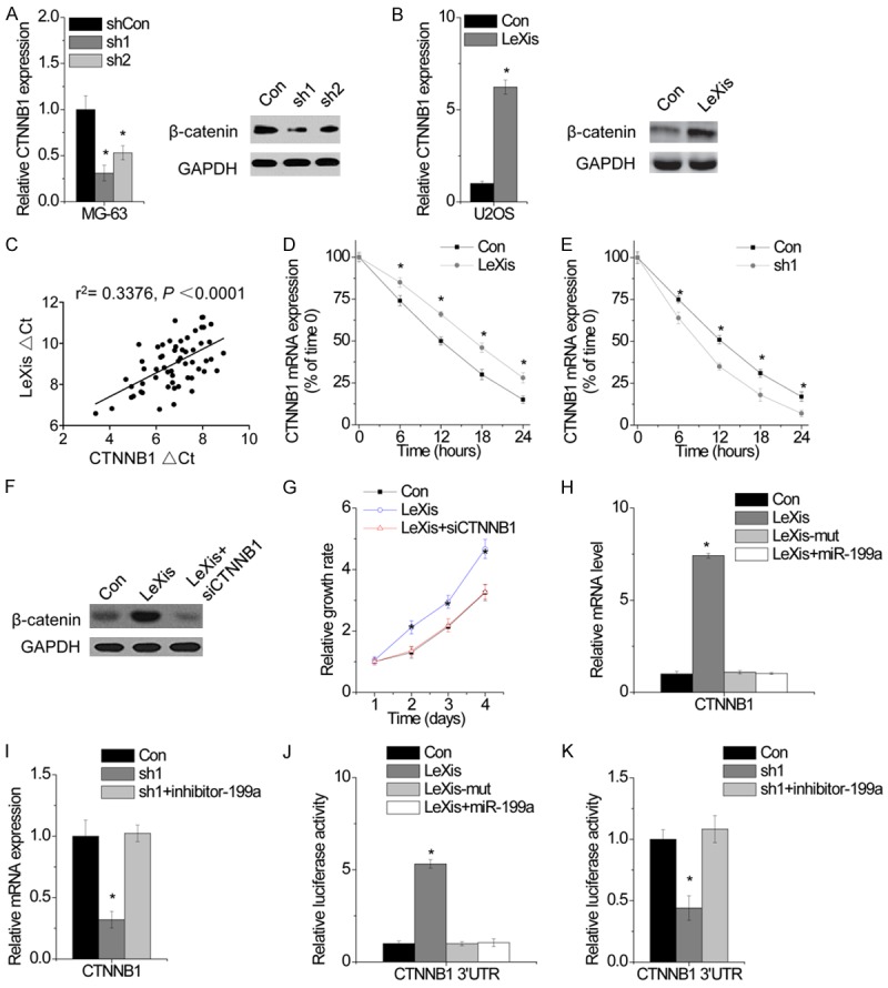
Figure 5.

LeXis functions as a ceRNA of β-catenin. A. The mRNA (left) and protein expression of CTNNB1 in MG-63 cells with or without LeXis knockdown was detected by qPCR and western blot, respectively. B. The mRNA (left) and protein expression of CTNNB1 in MG-63 cells with or without LeXis overexpression was detected by qPCR and western blot, respectively. C. The correlation between LeXis and CTNNB1 mRNA expression in 60 OS tissues. D. The stability of CTNNB1 over time was measured by qPCR relative to time 0 after blocking new RNA synthesis with α-amanitin (50 mM) in U2OS cells with LeXis overexpression and normalized to 18S rRNA (a product of RNA polymerase I that is unchanged by α-amanitin). E. The stability of CTNNB1 over time was measured by qPCR relative to time 0 after blocking new RNA synthesis with α-amanitin (50 mM) in MG-63 cells with LeXis knockdown and normalized to 18S rRNA (a product of RNA polymerase I that is unchanged by α-amanitin). F. siRNA against CTNNB1 was tranfected into U2OS cells with LeXis overexpression. G. The cell proliferation of U2OS cells expressing LeXis with and without CTNNB1 siRNA. H. The mRNA level of CTNNB1 expression in LeXis or LeXis-mut overexpressed U2OS cells cotransfected with miR-199a was analyzed by qPCR. I. The mRNA level of CTNNB1 expression in LeXis silenced MG-63 cells cotransfected with miR-199a inhibitor was analyzed by qPCR. J. The relative luciferase activity of CTNNB1 3’UTR in LeXis or LeXis-mut overexpressed U2OS cells cotransfected with miR-199a was analyzed by qPCR. K. The relative luciferase activity of CTNNB1 3’UTR in LeXis silenced MG-63 cells cotransfected with miR-199a inhibitor was analyzed. Data are shown as mean ± SD; *P < 0.05 (Student’s t test).
