Figure 2.

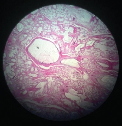
Pathologic feature shows hyperplastic and hypertrophied Bartholin’s glands, edema, chronic inflammatory infiltrate, focal dilation of ducts with squamous metaplasia, and increased number of acini.

Pathologic feature shows hyperplastic and hypertrophied Bartholin’s glands, edema, chronic inflammatory infiltrate, focal dilation of ducts with squamous metaplasia, and increased number of acini.