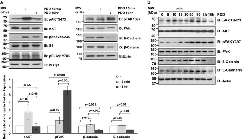
Figure 2.
Signaling events following PDD-induced TRPV4 activation. (a and b) 4T07 cells were untreated or treated with 4α-PDD at 10 μm for the indicated time points before subjecting the cell lysates to immunoblotting with the indicated antibodies. The protein bands in a were measured and quantified using Image-J software.

