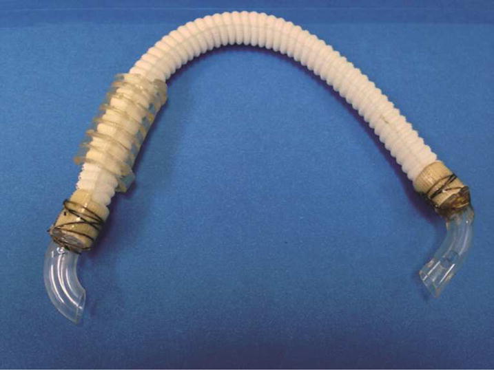
Figure 1.

Atrioventricular shunt using woven collagen embedded polyester and venous cannula. The shorter venous cannula (left side) is placed into the LA at the right superior pulmonary venous confluence. The longer perforated venous cannula (right side) is placed into the LV apex. Kinks are prevented using a coiled piece of cardiopulmonary bypass tubing for radial support.
