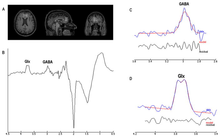
Fig. 2.
Proton Magnetic Resonance Spectroscopy (1H MRS). (A) a voxel (2.0 × 3.0 × 3.0 mm3) was placed into the ventro-medial prefrontal cortex by using T1-weighted image as anatomical reference; (B) representative raw GABA difference spectrum; (C, D) respectively report the representative GANNET-edited spectra (in blue) with estimated GABA and Glx models indicated in red. Residue was shown in black. (For interpretation of the references to color in this figure legend, the reader is referred to the web version of this article.)

